The Urinary System Pathology. Dr. Methaq Mueen د.ميثاق معين
|
|
|
- Chastity Marshall
- 5 years ago
- Views:
Transcription
1 The Urinary System Pathology Dr. Methaq Mueen د.ميثاق معين
2 objectives To Know normal anatomy physiology and histology of renal system To discuss Kidney pathology: congenital, cystic disease, Glmerular disease To know tubluointerstitial disease, UTI To discuss urinary bladder pathology

3 Structures of the Urinary System Kidneys Nephrons Renal Pelvis Ureters Urinary Bladder The Urethra
4 Functions of the Urinary System The urinary system performs many functions important to maintaining homeostasis Maintenance of water, salts, minerals and acids balance in the body Filters blood to remove urea and other waste products of metabolism from the bloodstream Urea the major waste product of protein metabolism Converts waste products and excess fluids into urine in the kidneys and excretes them from the body via the urinary bladder kidneys serve to convert more than 1700 L of blood per day into about 1 L of a highly concentrated fluid (urine). The kidney also serves as an endocrine organ, secreting such hormones as erythropoietin, renin, prostaglandins, and regulating vitamin D metabolism.

5 The Kidneys Filter blood constantly to remove waste products and excess water which are excreted as urine (95% water and 5% other wastes) 2 kidneys located retroperitoneally, one on each side of the vertebral column below the diaphragm consist of: Renal cortex The outer region of the kidney Contains over 1 million microscopic units called nephrons Medulla The inner region of the kidney Contains the urine collecting tubules

6 cotex cortex Medulla
7 Each human adult kidney weighs about 150 gm. Anatomically the ureter forms the pelvis (the dilated upper ureter) which is divided into 2 or 3 major calyces, each one giving 3 or 4 minor calyces. The kidney is divided into the cortex cm and medulla. The medulla consists of renal pyramids, the apices of which are called papillae, each related to a calyx.

8
9

10 1. Renal Vein 2. Renal Artery 3. Renal Calyx 4. Medullary Pyramid 5. Renal Cortex 6. Segmental Artery 7. InterlobAR Artery 8. Arcuate Artery interlobular 9. Arcuate Vein 10. Interlobar Vein 11. Segmental Vein 12. Renal Column 13. Renal Papillae 14. Renal Pelvis 15. Ureter
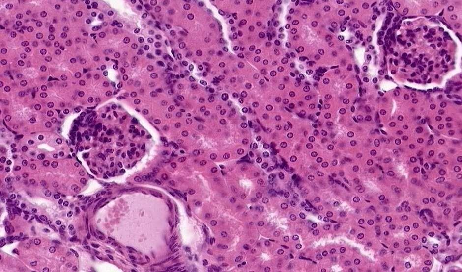
11 Nephrons The functional units of the kidneys Form urine by the process of filtration, reabsorption, and secretion Reabsorption: is the return of substances that were removed from filtration back to the bloodstream Each nephron contains a glomerulus Cluster of capillaries surrounded by a cup-shaped membrane called the Bowman s capsule
12 S.E.M. T.E.M.

13
14 How is Urine Made? Blood enters the kidneys through the renal artery and flows into the nephrons After being filtered by the capillaries of the glomerulus, the blood leaves the kidney through the renal vein Waste products that were filtered out of the blood remain behind in the kidney where they pass through urine-collecting tubules Urine is then transported to the renal pelvis and collected in preparation for entry into the ureters

15 The Renal Pelvis Funnel-shaped area in each kidney that is surrounded by the renal cortex and medulla Newly formed urine collects here before flowing to the ureters
16 The Ureters 2 narrow tubes (10-12 inches each) that transport urine from each kidney to the bladder Peristalsis moves urine down each ureter into the bladder
17 The Urinary Bladder Hollow muscular organ that is a reservoir or holding tank for urine before it is excreted from the body Located in the anterior portion of the pelvic cavity behind the pubic symphysis Lined with rugae that allow it to expand and contract Trigone smooth triangular area on the inner surface of the bladder located between the openings of the ureters and the urethra

18
19 KIDNEY
20 Kidney PATHOLOGY CONGENITAL MALFORMATION CYSTS GLOMERULAR DISEASES TUBULAR/INTERSTITIAL BLOOD VESSELS OBSTRUCTION TUMORS
21 CONGENITAL AGENESIS HYPOPLASIA ECTOPIC HORSESHOE
22 Congenital anomalies Agenesis of the kidney(absence): Bilateral agenesis is incompatible with life, seen in stillborn. Unilateral type is uncommon. The opposite kidney is usually enlarged as a result of compensatory hypertrophy. Hypoplasia(small size kidney) : Is failure of the kidney to develop to a normal size. This anomaly may occur bilaterally, resulting in renal failure and early childhood death. Unilateral cases are more common. The opposite kidney is also enlarged due to compensatory hypertrophy.
23 Renal (A) agenesis, and (B) hypoplasia. B

24 HYPOPLASIA
25 Congenital anomalies-cont Ectopic Kidneys: These kidneys lie within the pelvis. They are usually normal or slightly small in size. Because of their abnormal location can cause : kinking or tortuoisity of the ureters may cause some obstruction to urinary outflow. difficulty in labor in females. Misdiagnosis as pelvic tumors & abscesses. Have long renal artery that may have many complications like damage during surgery.

26 ECTOPIC (usually PELVIC)
27 Horseshoe kidneys: Fusion of lower poles of the kidneys that is continuous across the midline anterior to the great vessels. may cause many complications: 1. Partially obstructed the ureters, which result in hydronephrosis. 2. Recurrent UTI. 3. Stone formation.
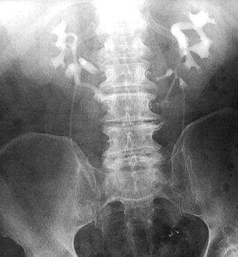

28 HORSESHOE
29 Cystic Diseases Of The Kidney They are heterogeneous group comprising hereditary, developmental and acquired disorders. They are important for several reasons; 1. They are reasonably common and often represent diagnostic problems for clinicians, radiologists and pathologists. 2. Some forms are major causes of chronic renal failure. 3. They can occasionally be confused with malignant tumors.
30 Classification of CYSTIC DISEASES Polycystic kidney : Autosomal DOMINANT (AD-ULTS) Autosomal RECESSIVE (CHILDREN) ACQUIRED SIMPLE (dialysis-associated) cystic disease.. Parasitic cysts (e.g. hydatid cyst).
31 Autosomal Dominant Polycystic Kidney Disease Multiple expanding cysts of both kidneys that ultimately destroy the intervening parenchyma. It affects roughly 1 of every 400 to 1000 live births and accounting for about 5-10% 0f cases of chronic renal failure. It can be caused by inheritance of at least two Autosomal dominant genes of high penetrance: 1. PKD 1, present on chromosome 16, mutant in 90% of cases. 2. PKD 2, present on chromosome 4, mutant
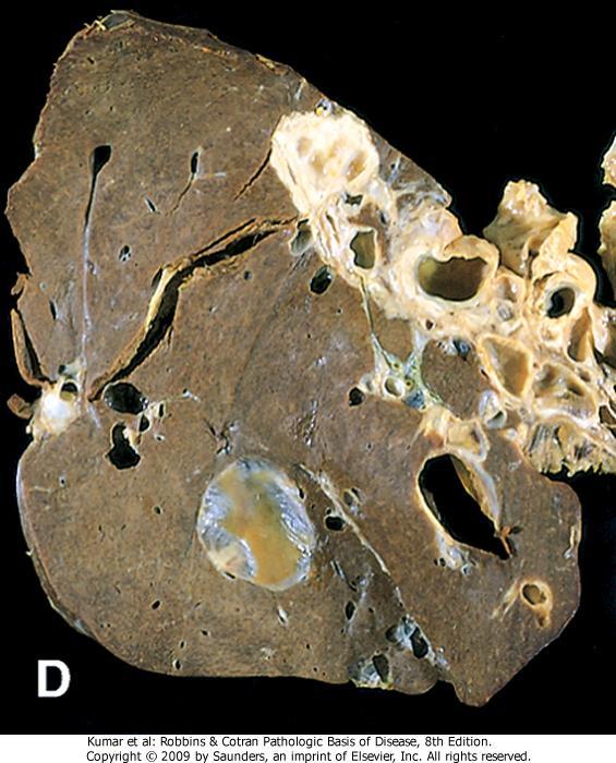
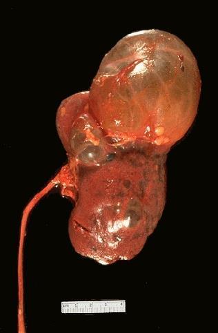
32 Gross: Large kidney (4 kg) of each kidney, & present as abdominal mass. Mass of cysts of varying sizes up to 3 to 4 cm in diameter without intervening parenchyma. Mic: Cysts are filled with fluid (clear, hemorrhagic). Cysts arise from tubules or collecting ducts. Often have atrophic lining. Superimposed infection is common..


33
34 Autosomal Dominant Polycystic Kidney Disease Clinical: Many of these patients remain asymptomatic until about the 4th or 5th decade of life when renal insufficiency occurs because the cysts initially involve only portions of the nephrones, so renal function is retained. Presentation (variable): flank pain, hypertension, hematuria, progressive renal failure Large lesions are palpable 40% have cystic disease of the liver (most common), spleen, pancreas, brain Berry aneurysms in circle of Willis and can cause death in about 4-10% of patients due to subarachnoid hemorrhage. Death due to uremia or hypertension 40% of patients die of coronary or hypertensive heart disease, 25% of infection, 15% of ruptured Berry aneurysm or hypertensive intracranial hemorrhage.


35

36
37
38 Autosomal Recessive (Childhood) Polycystic Kidney Disease Is rare anomaly due to mutation of gene PKHD1 on chromosome 6 Perinatal, neonatal, infantile and juvenile types. The first 2 are the most common In all cases are associated with liver cysts (congenital liver cirrhosis). Mic: Gross: bilateral Small cysts in the cortex & medulla (sponge like appearance). Elongated channels at right angles to the cortical surface. Uniform lining of cuboidal cells (Originate from collecting ducts).
39 Morphology The kidneys are enlarged (bilateral) and have a smooth external surface. On cut section, numerous small cysts in the cortex and medulla give the kidney a sponge-like appearance. Smooth, Small, Sponge The cysts are dilated channels perpendicular to the corticomedullary junction. Cysts originate from collective tubules and are lined by uniform cuboidal cells In almost all cases, the liver has cysts with portal fibrosis as well as proliferation of portal bile ducts. Clinical Features; serious manifestations are usually present at birth, and the young infant might succumb rapidly to renal failure. Patients, who survive infancy, may develop congenital hepatic fibrosis. Liver: epithelium lined cysts and proliferation of bile ducts


40


41
42 Acquired (Dialysis-Associated) Cystic Disease The kidneys of patients on chronic dialysis, sometimes exhibit numerous cortical and medullary cysts. The cysts measure 0.5-2cm, contain clear fluid, are lined by either hyperplastic or flattened tubular epithelium and often contain calcium oxalate crystals. Cause: They probably form as a result of tubular obstruction due to interstitial fibrosis or by oxalate crystals.
43 Acquired (Dialysis-Associated) Cystic Disease Most are asymptomatic, but sometimes the bleeding inside the cysts cause hematuria. The most important complication is the development of renal cell carcinoma in the walls of these cysts in about 7% of patients during 10 years period.

44
45 ACQUIRED (DIALYSIS)
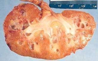
46 Simple Cysts Acquired, Incidental, very common These occur as single or multiple, usually cortical. The size range from 1-10 cm or more. They are translucent and filled with clear fluid. They are lined by a single layer of cuboidal or flattened epithelium. They are common postmortem findings. On occasion, hemorrhage into them may cause sudden pain, and calcification may be visible radiologically. The main importance of these cysts is in their differentiation from kidney tumors.

47
48
49 Renal diseases can be divided according to the parts of kidneys into 1. Glomerular diseases 2. Tubular diseases. 3. Interstitium diseases. 4. Blood vessels diseases.
50 This division is useful because a. The early manifestations of each group of diseases tend to be distinctive. b. These groups differ in their pathogenesis, for e.g., glomerular diseases are often immunologically mediated, whereas tubular and interstitial disorders are more likely to be caused by toxic or infectious agents. However, it should be noted that: 1. The interdependence of renal components translated into that damage to one component is almost always affects secondarily the others. 2. All forms of chronic renal disease tend ultimately to damage all four components of the kidney thus, eventuates in chronic renal failure (end-stage kidney disease ESKD).
51 GLOMERULAR DISEASES
52 Glomerular Diseases: They constitute some of the major problems in nephrology; in fact they are the most common causes of chronic renal failure in humans. Clinical manifestations of renal disease; Acute Nephritic Syndrome: characterized by Gross hematuria (macroscopic). Mild to moderate proteinuria. Edema. Hypertension Typical example is Poststreptococcal glomerulonephritis. 2. Nephrotic Syndrome: Heavy Proteinuria (> 3.5 gram of protein / 24hours). Hypoalbuminemia. Severe edema. Hyperlipidemia & lipiduria. 3. Asymptomatic hematuria & / or Proteinuria: Mild Glomerular abnormality.
53 4. Rapidly Progressive Glomerulonephritis: Loss of renal functions in a few days or weeks. Manifested by active urine sediment (hematuria, Dysmorphic RBC S, RBC S Casts). 5. Acute Renal failure: Oliguria (< 500 cc) or Anuria (no urine flow). Recent onset of Azotemia.(in Latin: nitrogen = azot) 6. Chronic Renal failure: Prolonged symptoms & signs of Uremia. Can be the end result of all chronic renal diseases. 7. Urinary tract infection: Characterized by Bacteruria & Pyuria ( bacteria and leukocytes in the urine). 8. Nephrolithiasis: Characterized by Renal colic. Important notes: Azotemia: is a biochemical abnormality that means elevation of blood urea nitrogen (BUN) and creatinine levels, and is related largely to a decreased glomerular filtration rate (GFR). Could be Prerenal Azotemia (Hypoperfusion of kidneys), renal (due to kidney diseases), Postrenal Azotemia (obstruction below the kidney). Uremia: characterized by Clinical Signs, Symptoms & Biochemical abnormalities. Renal damage (impair excretory, endocrine, & metabolic functions of kidneys). Uremic patients frequently manifest secondary involvement of the gastrointestinal system (e.g., uremic gastroenteritis), peripheral nerves (e.g., peripheral neuropathy), and heart (e.g., uremic fibrinous pericarditis).

54 Principal Systemic Manifestations of Chronic Kidney Disease and Uremia Fluid and Electrolytes

55 Clinical Presentations of GN


56

57
58 S.E.M. T.E.M.

59
60 One of important function of Glomerular wall is Selective Permeability (high permeable to H2o & small solutes) while completely impermeable to molecules of size & molecular charge of Albumin. The podocyte is decisive to the glomerular barrier function by providing a distal resistance to the flow of water and a barrier to the filtration of proteins. It is also largely responsible for synthesis of GBM components
61 Histologic alterations There are 5 basic tissue reactions 1. Increased glomerular cellularity a. Proliferation of mesangial or endothelial cells. b. Leukocyte infiltration, including neutrophils, monocytes, and, in some diseases, lymphocytes. c. Formation of crescents(proliferation of parietal epithelial cells. 2. Basement membrane thickening, best seen in sections stained with (PAS). By EM, it can be resolved as one of 2 alteration;
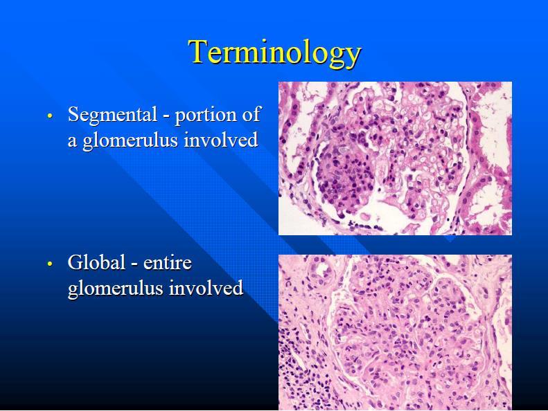
62 a. Deposition of amorphous electron dense material, of immune complexes, on the endothelial or epithelial side of basement membrane, or within the GBM itself. b. Thickening of the BM proper, as occurs in diabetic glomerulosclerosis. 3. Hyalinization and sclerosis, made up of plasma proteins and collagen material deposited exracellularly. 4. Additional alterations include; accumulation of lipids, fibrin or other metabolic materials. 5. Intraglomerular vascular thrombosis The histologic changes can be further subdivided into; 1. Diffuse 2. Focal. 3. Global. 4. Segmental. 5. Mesangial

63
64
65 Pathogenesis Although little is known about etiologic agents and triggering events, it is clear that immune mechanisms underlie most forms of primary glomerulopathies, and many of the secondary forms. Immune-mediated mechanisms; 1. Ab-mediated, a. Insitu immune complex deposition, In this form of injury, immune complexes are formed locally by antibodies that react with intrinsic tissue antigen or with extrinsic antigens "planted" in the glomerulus from the circulation. Fixed intrinsic antigens for e.g. (BM)like Goodpasterur syndrome (Ab against glomerular and pulmonary BM) The pattern of immune deposition by immunofluorescence microscopy is linear. Planted antigens (exogenous as infectious agent or drug and endogenous as DNA, immunoglobulins) The pattern of immune deposition by immunofluorescence microscopy is granular b. Circulating immune complex deposition Endogenous Ag (DNA, tumor) Exogenous Ag (infectious products)

66
67 Pathogenesis-cont 2. Cell-mediated. T cell-mediated injury may account for some cases of glomerulonephritis (GN) 3. Activation of alternative complement pathway.

68 Localization of immune complexes in the Glomerulus 1. Subepithelial humps, 2. Epimembranous deposits, 3. Subendothelial deposits, 4. Mesangial deposits, 5. Basement membrane. EN, endothelium; EP, epithelium; LD, lamina densa; LRE, lamina rara externa; LRI, lamina rara interna; MC, mesangial cell; MM, mesangial matrix.
69 GLOMERULAR DISEASES Primary Glomerulonephritis Acute diffuse proliferative GN Rapidly progressive GN Membranous GN Lipoid nephrosis (minimal change disease) Focal segmental glomerulosclerosis Membranoproliferative GN IgA Nephropathy Chronic GN Secondary (Systemic) Diseases Systemic lupus erythematosus Diabetes mellitus Amyloidosis Goodpasture s syndrome Polyarteritis nodosa Wagener s granulomatosis Henoch-Scholein purpura Bacterial endocarditis. Hereditary Disorders Alport s syndrome Fabry s disease
70 Nephritic Syndrome Injury.proliferation of the cells within the glomeruli(endothelium and mesangial cells), accompanied by leukocytic infiltrate. This inflammatory reaction injures the capillary walls(formation of holes in the basement membrane),. permitting escape of red cells into the urine, and induces hemodynamic changes that lead to a.reduction in the GFR. The reduced GFR is manifested clinically by oliguria, fluid retention, and azotemia. Hypertension is the result of both the fluid retention and excessive renin release Clinical Symptoms: Oliguria(due to decreased GFR) Azotemia (elevated creatnine and BUN) Hypertension (due to retention of salt and excessive renin secretion) mild-moderate Protinuria >150mg but <3.5g BUN/Creatinine level of >15 Hematuria best defined as red cell casts and RBC with dysmorphic membranes Commonly defined as smoky brown urine
 HYPERCELLULAR GLOMERULI INCREASED ENDOTHELIUM AND MESANGIUM IgG, IgM, C3 along GMB FOCALLY(Hump) 95% full")
71 ACUTE GLOMERULONEPHRITIS Nephritic Holes in the membrane Hematuria, Hypertension, Hardly any urin(oliguria), Azotemia in children following a strep infection POSTSTREPTOCOCCAL (old term) HYPERCELLULAR GLOMERULI INCREASED ENDOTHELIUM AND MESANGIUM IgG, IgM, C3 along GMB FOCALLY(Hump) 95% full recovery

72 RAPIDLY PROGRESSIVE GLOMERULONEPHRITIS Clinical definition, NOT a specific pathologic one CRESCENTIC Anti-GBM Ab IMMUN COMPLX Anti-Neut. Ab

73 Crescentic glomerulonephritis (PAS stain)

![5g/ day] Hypoalbuminemia [<3g/dl] Periorbital edema then Generalized edema Due to d plasma oncotic pressure Hyperlipiduria and Oval Fat Bodies](/docs-images/95/126048127/images/74-1.jpg "Hyperlidemia and Hypercholesterolemia due to loss of lipoproteins and alterations in liver production of lipoproteins Increase in Infections due to loss")
74 NEPHROTIC SYNDROME Syndrome of Glomerular dysfunction that is characterized by increased loss of proteins in the urine due to increased basement membrane permeability CLINICAL MANIFESTATIONS Massive proteinuria without hematuria [>3.5g/ day] Hypoalbuminemia [<3g/dl] Periorbital edema then Generalized edema Due to d plasma oncotic pressure Hyperlipiduria and Oval Fat Bodies Hyperlidemia and Hypercholesterolemia due to loss of lipoproteins and alterations in liver production of lipoproteins Increase in Infections due to loss of low weight globulins and complement Loss of anticoagulants hypercoagulable state
75 NEPHROTIC SYNDROME Only MASSIVE PROTEINURIA HYPOALBUMINEMIA EDEMA LIPIDEMIA/LIPIDURIA NUMEROUS CAUSES:
76 End
Functions of the kidney:
 Diseases of renal system : Normal anatomy of renal system : Each human adult kidney weighs about 150 gm, the ureter enters the kidney at the hilum, it dilates into a funnel-shaped cavity, the pelvis, from
Overview of glomerular diseases
 Overview of glomerular diseases *Endothelial cells are fenestrated each fenestra: 70-100nm in diameter Contractile, capable of proliferation, makes ECM & releases mediators *Glomerular basement membrane
Disorders of the kidney. Urine analysis. Nephrotic and nephritic syndrome.
 Disorders of the kidney. Urine analysis. Nephrotic and nephritic syndrome. Azotemia and Urinary Abnormalities Disturbances in urine volume oliguria, anuria, polyuria Abnormalities of urine sediment red
A clinical syndrome, composed mainly of:
 Nephritic syndrome We will discuss: 1)Nephritic syndrome: -Acute postinfectious (poststreptococcal) GN -IgA nephropathy -Hereditary nephritis 2)Rapidly progressive GN (RPGN) A clinical syndrome, composed
Alterations of Renal and Urinary Tract Function
 Alterations of Renal and Urinary Tract Function Chapter 29 Urinary Tract Obstruction Urinary tract obstruction is an interference with the flow of urine at any site along the urinary tract The obstruction
THE URINARY SYSTEM. The cases we will cover are:
 THE URINARY SYSTEM The focus of this week s lab will be pathology of the urinary system. Diseases of the kidney can be broken down into diseases that affect the glomeruli, tubules, interstitium, and blood
THE URINARY SYSTEM. The cases we will cover are:
 THE URINARY SYSTEM The focus of this week s lab will be pathology of the urinary system. Diseases of the kidney can be broken down into diseases that affect the glomeruli, tubules, interstitium, and blood
CYSTIC DISEASES of THE KIDNEY. Dr. Nisreen Abu Shahin
 CYSTIC DISEASES of THE KIDNEY Dr. Nisreen Abu Shahin 1 Types of cysts 1-Simple Cysts 2-Dialysis-associated acquired cysts 3-Autosomal Dominant (Adult) Polycystic Kidney Disease 4-Autosomal Recessive (Childhood)
Glomerular Pathology- 1 Nephrotic Syndrome. Dr. Nisreen Abu Shahin
 Glomerular Pathology- 1 Nephrotic Syndrome Dr. Nisreen Abu Shahin The Nephrotic Syndrome a clinical complex resulting from glomerular disease & includes the following: (1) massive proteinuria (3.5 gm /day
Glomerular pathology in systemic disease
 Glomerular pathology in systemic disease Lecture outline Lupus nephritis Diabetic nephropathy Glomerulonephritis Associated with Bacterial Endocarditis and Other Systemic Infections Henoch-Schonlein Purpura
Glomerular diseases mostly presenting with Nephritic syndrome
 Glomerular diseases mostly presenting with Nephritic syndrome 1 The Nephritic Syndrome Pathogenesis: proliferation of the cells in glomeruli & leukocytic infiltrate Injured capillary walls escape of RBCs
Urinary System. Genitourinary System (Part A-1) Module 10 -Chapter 11. Overview 1/4/2013
 Module 10 -Chapter 11. Overview 1/4/2013") Genitourinary System (Part A-1) Module 10 -Chapter 11 Overview Urinary system Kidneys Ureter Bladder Nephron Male reproductive system Testes Accessory Structures Susie Turner, M.D. 71/3/13 Major homeostatic
Genitourinary System (Part A-1) Module 10 -Chapter 11 Overview Urinary system Kidneys Ureter Bladder Nephron Male reproductive system Testes Accessory Structures Susie Turner, M.D. 71/3/13 Major homeostatic
Histopathology: Glomerulonephritis and other renal pathology
 Histopathology: Glomerulonephritis and other renal pathology These presentations are to help you identify basic histopathological features. They do not contain the additional factual information that you
Dr. Najla a Aldaoud. Omar Ayman Khasawneh
 Pathology 1 Congenital & Cystic diseases of the kidney Dr. Najla a Aldaoud Omar Ayman Khasawneh 1 P a g e Slides are included بسم هللا الرحمن الرحيم Today is our first pathology lectures, Dr Najla' will
URINARY SYSTEM I. Kidneys II. Nephron Unit and Urine Formation
 URINARY SYSTEM I. Kidneys A. Location and Structure 1. Retroperitoneal 2. Between T12 and L3 3. Rt. kidney slightly lower 4. Two bean shaped organs 5. Adrenal gland 6. Internal construction a. Renal cortex
Kidney Functions Removal of toxins, metabolic wastes, and excess ions from the blood Regulation of blood volume, chemical composition, and ph
 The Urinary System Urinary System Organs Kidneys are major excretory organs Urinary bladder is the temporary storage reservoir for urine Ureters transport urine from the kidneys to the bladder Urethra
Basic Urinary Tract Anatomy and Histology
 Basic Urinary Tract Anatomy and Histology The two kidneys are located in the retroperitoneum on either side of the vertebral bladder and the contraction of the detrusor muscle. Any mechanical barrier,
Glomerular pathology-2 Nephritic syndrome. Dr. Nisreen Abu Shahin
 Glomerular pathology-2 Nephritic syndrome Dr. Nisreen Abu Shahin 1 The Nephritic Syndrome Pathogenesis: inflammation proliferation of the cells in glomeruli & leukocytic infiltrate Injured capillary walls
Glomerulonephritis. Dr Rodney Itaki Anatomical Pathology Discipline.
 Glomerulonephritis Dr Rodney Itaki Anatomical Pathology Discipline. University of Papua New Guinea School of Medicine & Health Sciences Division of Pathology Gross anatomy Ref: Goggle Images Microanatomy
General Anatomy of Urinary System
 General Anatomy of Urinary System URINARY SYSTEM ORGANS Kidneys (2) Ureters (2) Urinary bladder Urethra KIDNEY FUNCTIONS Control blood volume and composition KIDNEY FUNCTIONS Filter blood plasma, eliminate
BCH 450 Biochemistry of Specialized Tissues
 BCH 450 Biochemistry of Specialized Tissues VII. Renal Structure, Function & Regulation Kidney Function 1. Regulate Extracellular fluid (ECF) (plasma and interstitial fluid) through formation of urine.
Urinary System Laboratory
 Urinary System Laboratory 1 Adrenal gland Organs of The Urinary System Renal artery and vein Kidney Ureter Urinary bladder Figure 26.1 2 Urethra Functions of the urinary system organs: Urethra expels urine
Urinary system. Urinary system
 INTRODUCTION. Several organs system Produce urine and excrete it from the body Maintenance of homeostasis. Components. two kidneys, produce urine; two ureters, carry urine to single urinary bladder for
Chapter 23. The Nephron. (functional unit of the kidney
 Chapter 23 The Nephron (functional unit of the kidney Renal capsule The Nephron Renal cortex Nephron Collecting duct Efferent arteriole Afferent arteriole (a) Renal corpuscle: Glomerular capsule Glomerulus
Nephrology - the study of the kidney. Urology - branch of medicine dealing with the male and female urinary systems and the male reproductive system
 Urinary System Nephrology - the study of the kidney Urology - branch of medicine dealing with the male and female urinary systems and the male reproductive system Functions of the Urinary System 1. Regulation
H I S T O L O G Y O F T H E U R I N A R Y S Y S T E M
 SCPA 602- Anatomical Basis For Pathological Study H I S T O L O G Y O F T H E U R I N A R Y S Y S T E M S O M P H O N G N A R K P I N I T, M. D. D E P A R T M E N T O F P A T H O B I O L O G Y F A C U
RENAL HISTOPATHOLOGY
 RENAL HISTOPATHOLOGY Peter McCue, M.D. Department of Pathology, Anatomy & Cell Biology Sidney Kimmel Medical College There are no conflicts of interest. 1 Goals and Objectives! Goals Provide introduction
Crescentic Glomerulonephritis (RPGN)
") Crescentic Glomerulonephritis (RPGN) Background Rapidly progressive glomerulonephritis (RPGN) is defined as any glomerular disease characterized by extensive crescents (usually >50%) as the principal histologic
Crescentic Glomerulonephritis (RPGN) Background Rapidly progressive glomerulonephritis (RPGN) is defined as any glomerular disease characterized by extensive crescents (usually >50%) as the principal histologic
Lab Activity 31. Anatomy of the Urinary System. Portland Community College BI 233
 Lab Activity 31 Anatomy of the Urinary System Portland Community College BI 233 Urinary System Organs Kidneys Urinary bladder: provides a temporary storage reservoir for urine Paired ureters: transport
CHAPTER 25 URINARY. Urinary system. Kidneys 2 Ureters 2 Urinary Bladder 1 Urethra 1. functions
 CHAPTER 25 URINARY Kidneys 2 Ureters 2 Urinary Bladder 1 Urethra 1 fluid waste elimination secretion of wastes control blood volume and BP control blood ph electrolyte levels RBC levels hormone production
Human Urogenital System 26-1
 Human Urogenital System 26-1 Urogenital System Functions Filtering of blood, Removal of wastes and metabolites Regulation of blood volume and composition concentration of blood solutes ph of extracellular
Advanced Concept of Nursing- II UNIT-VI Advance Nursing Management of Genitourinary (GU) Diseases.
 Diseases.") In The Name of God (A PROJECT OF NEW LIFE COLLEGE OF NURSING KARACHI) Advanced Concept of Nursing- II UNIT-VI Advance Nursing Management of Genitourinary (GU) Diseases. Shahzad Bashir RN, BScN, DCHN,MScN
In The Name of God (A PROJECT OF NEW LIFE COLLEGE OF NURSING KARACHI) Advanced Concept of Nursing- II UNIT-VI Advance Nursing Management of Genitourinary (GU) Diseases. Shahzad Bashir RN, BScN, DCHN,MScN
Figure 26.1 An Introduction to the Urinary System
 Chapter 26 Figure 26.1 An Introduction to the Urinary System Components of the Urinary System Kidney Produces urine Ureter Transports urine toward the urinary bladder Urinary Bladder Temporarily stores
The Urinary System. Medical Assisting Third Edition. Booth, Whicker, Wyman, Pugh, Thompson The McGraw-Hill Companies, Inc. All rights reserved
 The Urinary System PowerPoint presentation to accompany: Medical Assisting Third Edition Booth, Whicker, Wyman, Pugh, Thompson 30-2 Learning Outcomes 30.1 Describe the structure, location, and functions
Nephritic vs. Nephrotic Syndrome
 Page 1 of 18 Nephritic vs. Nephrotic Syndrome Terminology: Glomerulus: A network of blood capillaries contained within the cuplike end (Bowman s capsule) of a nephron. Glomerular filtration rate: The rate
Urinary Physiology. Chapter 17 Outline. Kidney Function. Chapter 17
 Urinary Physiology Chapter 17 Chapter 17 Outline Structure and Function of the Kidney Glomerular Filtration Reabsorption of Salt and Water Renal Plasma Clearance Renal Control of Electrolyte and Acid-Base
Urinary System Organization. Urinary System Organization. The Kidneys. The Components of the Urinary System
 Urinary System Organization The Golden Rule: The Job of The Urinary System is to Maintain the Composition and Volume of ECF remember this & all else will fall in place! Functions of the Urinary System
PATTERNS OF RENAL INJURY
 PATTERNS OF RENAL INJURY Normal glomerulus podocyte Glomerular capillaries electron micrograph THE CLINICAL SYNDROMES 1. The Nephrotic Syndrome 2. The Acute Nephritic Syndrome 3. Rapidly Progressive Glomerulonephritis
Histology Urinary system
 Histology Urinary system Urinary system Composed of two kidneys, two ureters, the urinary bladder, and the urethra, the urinary system plays a critical role in: 1- Blood filtration,(filtration of cellular
Dr Ian Roberts Oxford. Oxford Pathology Course 2010 for FRCPath Illustration-Cellular Pathology. Oxford Radcliffe NHS Trust
 Dr Ian Roberts Oxford Oxford Pathology Course 2010 for FRCPath Present the basic diagnostic features of the commonest conditions causing proteinuria & haematuria Highlight diagnostic pitfalls Nephrotic
URINARY SYSTEM CHAPTER 28 I ANATOMY OF THE URINARY SYSTEM. Student Name
 Student Name CHAPTER 28 URINARY SYSTEM L iving produces wastes. Wherever people live or work or play, wastes accumulate. To keep these areas healthy, there must be a method of disposing of these wastes
19. RENAL PHYSIOLOGY ROLE OF THE URINARY SYSTEM THE URINARY SYSTEM. Components and function. V BS 122 Physiology II 151 Class of 2011
 19. RENAL PHYSIOLOGY THE URINARY SYSTEM Components and function The urinary system is composed of two kidneys, the functionally filtering apparatus, which connect through two tubular structures called
Urinary System. consists of the kidneys, ureters, urinary bladder and urethra
 Urinary System 1 Urinary System consists of the kidneys, ureters, urinary bladder and urethra 2 Location of Kidneys The kidneys which are positioned retroperitoneally lie on either side of the vertebral
URINARY SYSTEM. These organs lie posterior or inferior to the. (membrane).
.") URINARY SYSTEM I. INTRODUCTION Each kidney is made up of about a million tiny tubules called nephrons. Each nephron individually filters the blood and makes urine and it does the job completely, from start
URINARY SYSTEM I. INTRODUCTION Each kidney is made up of about a million tiny tubules called nephrons. Each nephron individually filters the blood and makes urine and it does the job completely, from start
Dr. Rai Muhammad Asghar Head of Paediatric Department BBH Rawalpindi
 Dr. Rai Muhammad Asghar Head of Paediatric Department BBH Rawalpindi Acute Post streptococcal Glomerulonephritis Sudden onset of Gross hematuria Edema Hypertension Renal insufficiency Cause of AGN Post
URINARY SYSTEM. Urinary System
 URINARY SYSTEM Urinary System Kidney Functions Excretion Regulation of blood volume and pressure Regulation of electrolyte and ph levels Kidney Structure Gross Anatomy Fibrous Capsule Renal Cortex Renal
Urinary System. Unit 6.12 (6 th Edition) Chapter 7.12 (7 th Edition)
 Chapter 7.12 (7 th Edition)") Urinary System Unit 6.12 (6 th Edition) Chapter 7.12 (7 th Edition) 1 Learning Objectives Identify the major organs of the urinary system and their functions. Explain the major functions of the kidneys.
Urinary System Unit 6.12 (6 th Edition) Chapter 7.12 (7 th Edition) 1 Learning Objectives Identify the major organs of the urinary system and their functions. Explain the major functions of the kidneys.
Year 2004 Paper one: Questions supplied by Megan
 QUESTION 53 Endothelial cell pathology on renal biopsy is most characteristic of which one of the following diagnoses? A. Pre-eclampsia B. Haemolytic uraemic syndrome C. Lupus nephritis D. Immunoglobulin
Copyright 2003 Pearson Education, Inc. publishing as Benjamin Cummings. Dr. Nabil Khouri
 Dr. Nabil Khouri Objectives: General objectives: - to identify the kidney s structures, function and location - to analyze the relationship between microscopic structure and function Specific objectives:
The topic of normal vascular and glomerular anatomy is introduced
 Normal Vascular and Glomerular Anatomy Arthur H. Cohen Richard J. Glassock The topic of normal vascular and glomerular anatomy is introduced here to serve as a reference point for later illustrations of
Cystic Renal Disease, for USMLE Step One. Howard J. Sachs, MD
 Cystic Renal Disease, for USMLE Step One Howard J. Sachs, MD www.12daysinmarch.com The Major Players Medullary Sponge Kidney (MSK) Polycystic Kidney Disease (PKD) Autosomal Recessive: Childhood Autosomal
Ch17-18 Urinary System
 Ch17-18 Urinary System Main Function: Filter the blood Other Functions: maintain purity and consistency of internal fluids eliminates nitrogenous wastes, toxins, and drugs from the body regulates blood
RNPDC CCNP Anatomy and Physiology: Renal System Pre-Quiz 2015
 RNPDC CCNP Anatomy and Physiology: Renal System Pre-Quiz 2015 1. In which abdominal cavity do the kidneys lie? a) Peritoneum. b) Anteperitoneal. c) Retroperitoneal. d) Parietal peritoneal 2. What is the
Elevated Serum Creatinine, a simplified approach
 Elevated Serum Creatinine, a simplified approach Primary Care Update Creighton University School of Medicine. April 27 th, 2018 Disclosure Slide I have no disclosures and have no conflicts with this presentation.
PHGY210 Renal Physiology
 PHGY210 Renal Physiology Tomoko Takano, MD, PhD *Associate Professor of Medicine and Physiology McGill University *Nephrologist, McGill University Health Centre Lecture plan Lecture 1: Anatomy, basics
April 08, biology 2201 ch 11.3 excretion.notebook. Biology The Excretory System. Apr 13 9:14 PM EXCRETORY SYSTEM.
 Biology 2201 11.3 The Excretory System EXCRETORY SYSTEM 1 Excretory System How does the excretory system maintain homeostasis? It regulates heat, water, salt, acid base concentrations and metabolite concentrations
URINARY SYSTEM ANATOMY
 URINARY SYSTEM ANATOMY Adapted from Human Anatomy & Physiology Marieb and Hoehn (9 th ed.) OVERVIEW Metabolism of nutrients by the body produces wastes that must be removed from the body. Although excretory
Tubulointerstitial Renal Disease. Anna Vinnikova, MD Division of Nephrology
 Tubulointerstitial Renal Disease Anna Vinnikova, MD Division of Nephrology Part I: Cystic Renal Disease www.pathguy.com Simple cysts Simple cysts May be multiple Usually 1 5cm, may be bigger Translucent,
URINARY SYSTEM ANATOMY PART
 URINARY SYSTEM ANATOMY PART 1 DANIL HAMMOUDI.MD Urinary System Composed of kidneys, ureters, urinary bladder, and urethra Eliminates nitrogenous wastes from the body Regulates water, electrolyte, and ph
The functions of the kidney:
 The functions of the kidney: After reading this lecture you should be able to.. 1. List the main functions of the kidney. 2. Know the basic physiological anatomy of the kidney and the nephron 3. Describe
Human Anatomy Unit 3 URINARY SYSTEM
 Human Anatomy Unit 3 URINARY SYSTEM In Anatomy Today Components Kidneys Ureters Urinary bladder Urethra Functions Storage of urine Bladder stores up to 1 L of urine Excretion of urine Transport of urine
Urinary System kidneys, ureters, bladder & urethra
 Urinary System kidneys, ureters, bladder & urethra Kidney Function Filters blood removes waste products conserves salts, glucose, proteins, nutrients and water Produces urine Endocrine functions regulates
network of thin-walled capillaries closely surrounded by a pear-shaped epithelial membrane called the Bowman s capsule
 Renal Terminology Renal-Root Words & Combining Forms calyx cortex glomerul/o medulla nephr/o pyel/o py/o ur/o ren/o cuplike division of the kidney outer layer of kidney glomerula inner or central portion
Dr P Sigwadi 30 May 2012
 Dr P Sigwadi 30 May 2012 Introduction Haematuria Positive blood on urine dipstick 5 red blood cells/ microliter of urine Prevalence Gross haematuria ( macroscopic) 0.13 % Microscopic- 1.5% Haematuria +
ESRD Dialysis Prevalence - One Year Statistics
 Age Group IL Other Total 00-04 12 1 13 05-09 5 2 7 10-14 15 1 16 15-19 55 2 57 20-24 170 10 180 25-29 269 14 283 30-34 381 9 390 35-39 583 14 597 40-44 871 20 891 45-49 1,119 20 1,139 50-54 1,505 35 1,540
NEPHRITIC SYNDROME. By Dr Mai inbiek
 NEPHRITIC SYNDROME By Dr Mai inbiek Nephritic Syndrome The nephritic Syndrome is a clinical complex, usually of acute onset. Is caused by inflammatory lesions of glomeruli. Characterized by; 1) Hematuria
Renal. Pathology. Kris%ne Kra*s, M.D.
 Renal Pathology Kris%ne Kra*s, M.D. Renal Pathology Outline Introductory stuff Glomerular diseases Tubular and inters%%al diseases Diseases involving blood vessels Cys%c diseases Tumors Renal Pathology
Urinary System Multiple Choice Practice Test. c. Kidneys have three protective layers d. The adrenal gland is located deep within the kidney
 Urinary System Multiple Choice Practice Test 1. Which of the following is a function of the urinary system? a. Regulates water b. Regulates balance of acids, bases, and electrolytes c. Filters waste from
Urinary System BIO 250. Waste Products of Metabolism Urea Carbon dioxide Inorganic salts Water Heat. Routes of Waste Elimination
 Urinary System BIO 250 Waste Products of Metabolism Urea Carbon dioxide Inorganic salts Water Heat Routes of Waste Elimination Skin: Variable amounts of heat, salts, and water; small amounts of urea and
28/04/2013 LEARNING OUTCOME C13 URINARY SYSTEM STUDENT ACHIEVEMENT INDICATORS STUDENT ACHIEVEMENT INDICATORS URINARY SYSTEM & EXCRETION
 LEARNING OUTCOME C13 Analyse the functional interrelationships of the structures of the urinary system Learning Outcome C13 URINARY SYSTEM STUDENT ACHIEVEMENT INDICATORS Students who have fully met this
AN APPROACH TO HEMATURIA. Dr Saima Ali
 AN APPROACH TO HEMATURIA Dr Saima Ali Definition Microscopic hematuria hematuria is defined as the presence of 5 or more RBCs per high-power field in 3 of 3 consecutive centrifuged specimens obtained at
Renal pathophysiology.
 Renal pathophysiology basa.konecna@gmail.com Outline Intro basic structure & physiology Nephrotic syndrome Nephritic syndrome Acute renal failure Chronic kidney disease Gross structure and location Kidney
Renal Pathology 1: Glomerulus. With many thanks to Elizabeth Angus PhD for EM photographs
 Renal Pathology 1: Glomerulus With many thanks to Elizabeth Angus PhD for EM photographs Anatomy of the Kidney http://www.yalemedicalgroup.org/stw/page.asp?pageid=stw028980 The Nephron http://www.beltina.org/health-dictionary/nephron-function-kidney-definition.html
The Urinary System 15PART A. PowerPoint Lecture Slide Presentation by Patty Bostwick-Taylor, Florence-Darlington Technical College
 PowerPoint Lecture Slide Presentation by Patty Bostwick-Taylor, Florence-Darlington Technical College The Urinary System 15PART A Functions of the Urinary System Elimination of waste products Nitrogenous
GLOMERULAR TUBULAR/INTERSTITIAL BLOOD VESSELS
 KIDNEY RENAL PATHOLOGY NORMAL CONGENITAL CYSTS GLOMERULAR TUBULAR/INTERSTITIAL BLOOD VESSELS OBSTRUCTION TUMORS 1. Renal Vein 2. Renal Artery 3. Renal Calyx 4. Medullary Pyramid 5. Renal Cortex 6. Segmental
Surgical Pathology Report
 Louisiana State University Health Sciences Center Department of Pathology Shreveport, Louisiana Accession #: Collected: Received: Reported: 6/1/2012 09:18 6/2/2012 09:02 6/2/2012 Patient Name: Med. Rec.
Urinary bladder provides a temporary storage reservoir for urine
 Urinary System Organs Kidney Filters blood, allowing toxins, metabolic wastes, and excess ions to leave the body in urine Urinary bladder provides a temporary storage reservoir for urine Paired ureters
The Urinary System PART A
 15 The Urinary System PART A PowerPoint Lecture Slide Presentation by Jerry L. Cook, Sam Houston University ESSENTIALS OF HUMAN ANATOMY & PHYSIOLOGY EIGHTH EDITION ELAINE N. MARIEB Functions of the Urinary
organs of the urinary system
 organs of the urinary system Kidneys (2) bean-shaped, fist-sized organ where urine is formed. Lie on either sides of the vertebral column, in a depression beneath peritoneum and protected by lower ribs
Bio 322 Human Anatomy Objectives for the laboratory exercise Urinary System Filtration Reabsorption Secretion Concentration
 Bio 322 Human Anatomy Objectives for the laboratory exercise Urinary System Required reading before beginning this lab: Saladin, KS: Human Anatomy 5 th ed (2017) Chapter 25 For this lab you will use parts
A. Correct! Flushing acids from the system will assist in re-establishing the acid-base equilibrium in the blood.
 OAT Biology - Problem Drill 16: The Urinary System Question No. 1 of 10 1. Which of the following would solve a drop in blood ph? Question #01 (A) Decreased retention of acids. (B) Increased excretion
The Excretory System
 The Excretory System The excretory system The excretory system includes the skin, lungs and kidneys which all release metabolic wastes from the body. The kidneys, skin and the lungs are the principle organs
Waste. Urinary System Anatomy Urinary Section pages 5-8. Urinary System. Urinary System. Nitrogenous Wastes. Nitrogenous Wastes 4/22/2016
 Waste Urinary System Anatomy Urinary Section pages 5-8 Metabolism produces waste products What is the primary waste product of cellular respiration? How does the body dispose of it? Urinary System Urinary
Urinary System kidneys, ureters, bladder & urethra
 Urinary System kidneys, ureters, bladder & urethra Filters blood removes waste products conserves salts, glucose, proteins, nutrients and water Produces urine Kidney Function Endocrine functions regulates
BIOL2030 Human A & P II -- Exam 6
 BIOL2030 Human A & P II -- Exam 6 Name: 1. The kidney functions in A. preventing blood loss. C. synthesis of vitamin E. E. making ADH. B. white blood cell production. D. excretion of metabolic wastes.
Day 1 Bell Work We will be discussing one of FIVE excretory organs in the human body. We have already studied four of them. The kidneys are considered
 URINARY SYSTEM 1 Day 1 Bell Work We will be discussing one of FIVE excretory organs in the human body. We have already studied four of them. The kidneys are considered the main organ in the excretory system.
1. Urinary System, General
 S T U D Y G U I D E 16 1. Urinary System, General a. Label the figure by placing the numbers of the structures in the spaces by the correct labels. 7 Aorta 6 Kidney 8 Ureter 2 Inferior vena cava 4 Renal
I. Anatomy of the Urinary System A. Kidneys 1. Right lower than Left* 2. Retroperitoneal 3. Layers that secure kidneys in the abdominal cavity a.
 I. Anatomy of the Urinary System A. Kidneys 1. Right lower than Left* 2. Retroperitoneal 3. Layers that secure kidneys in the abdominal cavity a. Renal fascia b. Perinephric fat (Adipose) capsule c. Fibrous
SHORT ANSWER. Write the word or phrase that best completes each statement or answers the question.
 Exam Name SHORT ANSWER. Write the word or phrase that best completes each statement or answers the question. Figure 25.1 Using Figure 25.1, match the following: 1) Glomerulus. 2) Afferent arteriole. 3)
A. Incorrect! The urinary system is involved in the regulation of blood ph. B. Correct! The urinary system is involved in the synthesis of vitamin D.
 Human Anatomy - Problem Drill 22: The Urinary System Question No. 1 of 10 1. Which of the following statements about the functions of the urinary system is not correct? Question #01 (A) The urinary system
EXCRETION IN HUMANS 31 JULY 2013
 EXCRETION IN HUMANS 31 JULY 2013 Lesson Description In this lesson we: Discuss organs of excretion Look at the structure of the urinary system Look at the structure and functioning of the kidney Discuss
ACUTE GLOMERULONEPHRITIS. IAP UG Teaching slides
 ACUTE GLOMERULONEPHRITIS 1 Definition Etiology Pathology/pathogenesis Risk factors Clinical Presentation Investigation Differential Diagnosis Management Outcome/Prognosis Indication for Renal Biopsy Summary
Chapter 14 The Urinary System. Anatomy
 Chapter 14 The Urinary System Anatomy 14.1 Overview Role in removal of wastes to maintain homeostasis Acts as filtering system of the blood Produces urine Removes wastes, maintains ph, electrolyte composition,
The Urinary System. Copyright 2003 Pearson Education, Inc. publishing as Benjamin Cummings
 The Urinary System Functions of the Urinary System Elimination of waste products Nitrogenous wastes Toxins Drugs Functions of the Urinary System Regulate aspects of homeostasis Water balance Electrolytes
Chapter 25: Urinary System
 Chapter 25: Urinary System I. Kidney anatomy: retroperitoneal from 12 th thoracic to 3 rd lumbar area A. External anatomy: hilus is the indentation 1. Adrenal gland: in the fat at the superior end of each
Functions of the Urinary System
 The Urinary System Functions of the Urinary System Elimination of waste products Nitrogenous wastes Toxins Drugs Regulate aspects of homeostasis Water balance Electrolytes Acid-base balance in the blood
HIHIM 409 7/26/2009. Kidney and Nephron. Fermamdo Vega, M.D. 1
 Function of the Kidneys Nephrology Fernando Vega, M.D. Seattle Healing Arts Center Remove Wastes Regulate Blood Pressure Regulate Blood Volume Regulates Electrolytes Converts Vitamin D to active form Produces
Outline Urinary System. Urinary System and Excretion. Urine. Urinary System. I. Function II. Organs of the urinary system
 Outline Urinary System Urinary System and Excretion Bio105 Chapter 16 Renal will be on the Final only. I. Function II. Organs of the urinary system A. Kidneys 1. Function 2. Structure III. Disorders of
The Urinary S. (Chp. 10) & Excretion. What are the functions of the urinary system? Maintenance of water-salt and acidbase
 & Excretion. What are the functions of the urinary system? Maintenance of water-salt and acidbase") 10.1 Urinary system The Urinary S. (Chp. 10) & Excretion 10.1 Urinary system What are the functions of the urinary system? 1. Excretion of metabolic wastes (urea, uric acid & creatinine) 1. Maintenance
10.1 Urinary system The Urinary S. (Chp. 10) & Excretion 10.1 Urinary system What are the functions of the urinary system? 1. Excretion of metabolic wastes (urea, uric acid & creatinine) 1. Maintenance
Urinary System. Analyze the Anatomy and Physiology of the urinary system
 Urinary System Analyze the Anatomy and Physiology of the urinary system Kidney Bean-shaped Located between peritoneum and the back muscles (retroperitoneal) Renal pelvis funnelshaped structure at the beginning